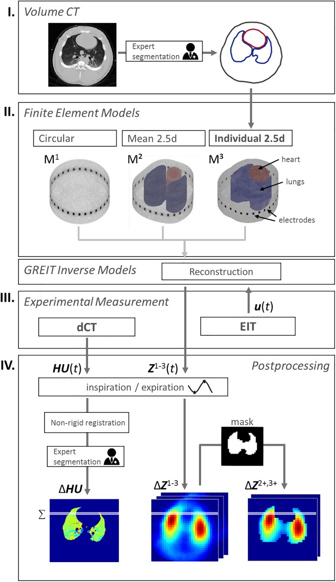
Fig 1. Acquisition and processing procedure.
I) Volume CTs were recorded for each pig and the contours of thorax, lungs and heart were extracted by radiologists. II) Finite element models were created using no prior information (M1), averaged contours (M2) or individual contours of each animal (M3). These models and optimized reconstruction settings were utilized to calculate the reconstruction matrix of EIT. III) Experimental measurement of 4DCT and EIT as well as reconstruction of EIT image series Z1-3(t) and (IV) extraction of tidal volume images and their ventilation profiles.

