Figure 4.
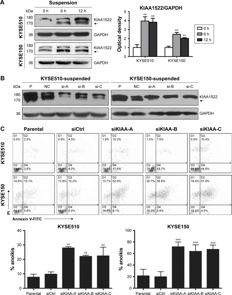
KIAA1522 depletion enhances anoikis in ESCC cells.
Notes: (A) KYSE510 and KYSE150 cells were cultured in suspension for the indicated times, and the KIAA1522 protein levels were detected by Western blotting. Relative ratios of absorbance for KIAA1522 to GAPDH were plotted. The data are presented as the mean ± SEM. **P<0.01. The arrows in images A and B indicate the position of KIAA1522 specific band. (B–D) KYSE150 and KYSE510 cells were transiently transfected with KIAA1522 siRNA for 48 h and then cultured on polyHEMA-coated dishes for 20 h. (B) KIAA1522 expression was determined by Western blot. (C) Anoikis was measured by Annexin V-FITC/PI staining and flow cytometry analysis. Representative results are shown (upper panel), and the percentages of apoptotic cells were plotted (lower panel). The data are presented as the mean ± SEM. **P<0.01, ***P<0.001.
Abbreviations: Ctrl, control; ESCC, esophageal squamous cell carcinoma; FITC, fluorescein isothiocyanate; GAPDH, glyceraldehyde 3-phosphate dehydrogenase; NC, non-silencing; P, parental; PI, propidium iodide; polyHEMA, polyhydroxylethylmethacrylate; si, siRNA; si-A, siKIAA-A; si-B, siKIAA-B; si-c, siKIAA-C.

