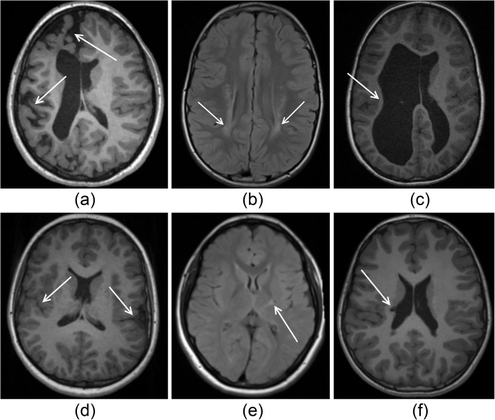
Fig 1. Illustrations of severe injury (top row) and subtle injury (bottom row) in cases of cortical malformations (first column), white/grey matter lesions (middle column) and ventricular enlargement (last column).
(A) WM atrophy with corresponding ventricular enlargement is presented with abnormal sulcal depth predominantly in the frontal and occipital lobes. (B) WM lesions resulting from periventricular leukomalacia are shown as local regions of high intensity. (C) The consequences of a periventricular haemorrhagic infarction leading to a severe loss of WM and secondary enlargement of the lateral ventricles is shown, particularly on the left hemisphere. (D) Bilateral perisylvian polymicrogyria is shown, which are visible as excessive numbers of small gyri. (E) Gliosis in a small location in the posterior limb of the internal capsule is shown. (F) Periventricular cystic lesion in the right ventricular cella media is shown, leading to a small region of ventricular enlargement on the lateral side of the ventricle.

