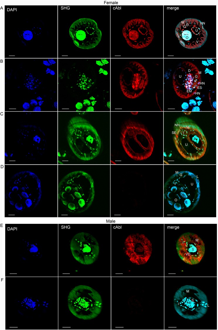
Fig 1. Expression of c-Abl homologue in B. malayi adults.
(A-C) Adult females and (E) adult males were exposed to polyclonal anti-c-Abl rabbit antibody or (D, F) isotype control rabbit IgG and counterstained with Alexa Fluor 594 labeled goat anti-rabbit IgG. High levels of expression are seen throughout the hypodermis (including hypodermal cords), gastrointestinal tract, uteri (A, female), and vas deferens (E, male). Scale bars are 20μm. O, fertilized ova; U, uterus; M, muscle; L, lateral chord; EM, early morula stage; P, early pretzel stage; ES, excretory secretory canal; S, spermatids; VD, Vas Deferens; D, dorsal cord; V, ventral cord; HN, hypodermal nuclei; I, intestine. Images are representative of at least 6 immunofluorescent microscopy experiments.

