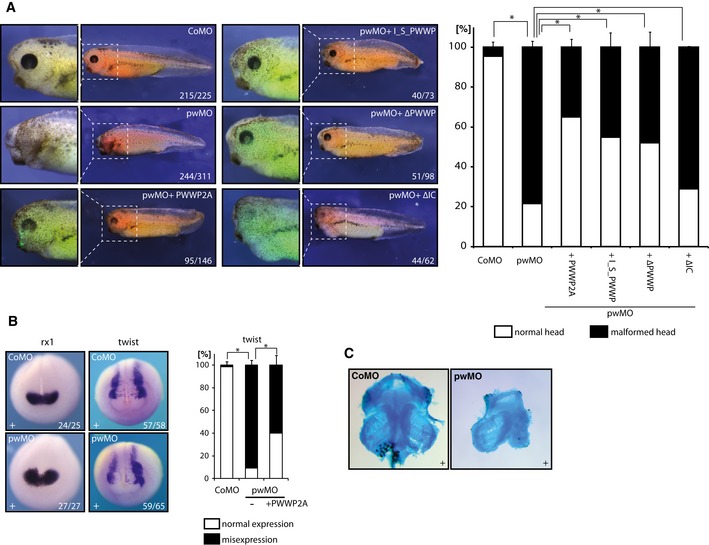
Figure 8. PWWP2A is essential for Xenopus eye, head, and brain development.

- Left side: One‐cell of two‐cell stage Xenopus laevis embryos was injected with either control (CoMO) or pwwp2a‐specific (pwMO) morpholino. For rescue experiments, pwMO morphant embryos were coinjected with mRNAs encoding either GFP‐tagged full‐length or variant human PWWP2A proteins, as indicated. Injected body sites were identified by Alexa 594 red fluorescence, while GFP fluorescence indicates synthesis of coinjected human PWWP2A protein variants. Panels display side views of representative embryos from the indicated conditions; numbers indicate penetrance of the major morphological phenotype over total embryos inspected. Right side: Quantification of the percentage of the observed phenotype, that is, malformation of head structures and eyes. Error bars indicate SEM of at least three independent biological replicates (see also Table EV1). *P ≤ 0.05 (Student's t‐test, two‐sided, unpaired).
- Whole‐mount RNA in situ hybridization assays with probes against rx1 (anterior view) and twist (dorsoanterior view) in either CoMO‐ or pwMO‐injected embryos at neurula stage. The rx1 gene is induced normally; the cranial neural crest marker twist is diminished on the pwMO‐injected side (91% affected), which is partially restored by coinjection of full‐length PWWP2A mRNA (60% affected). Numbers indicate penetrance of the depicted molecular phenotype over total embryos inspected. Right: Quantification of the percentage of misexpression of twist mRNA in controls compared to pwMO morphant and rescue condition. Error bars indicate SEM of three independent biological replicates. *P ≤ 0.05 (Student's t‐test, two‐sided, unpaired).
- Representative images of dissected facial cartilage from CoMO‐ or pwMO‐injected embryos visualized by Alcian blue staining. (+) indicates injected body side.
