
Figure 2. Negative stain EM structure of RERdj3WT reveals the native tetramer.
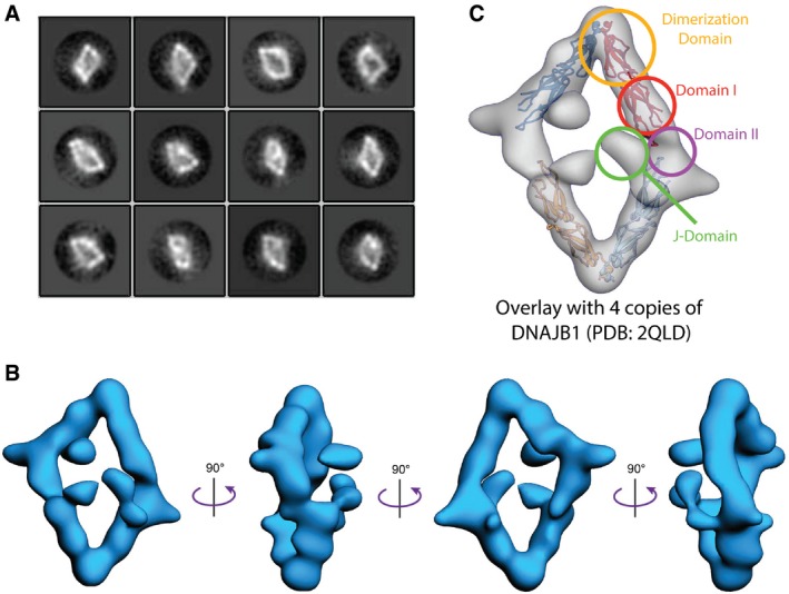
- 2D reference‐free class averages of negative stained RERdj3WT complexes show a diamond‐like shape.
- A 19 Å resolution structure of the RERdj3 tetramer is shown in 4 orthogonal views, displaying the flat, diamond‐shaped organization of the tetramer. The EM map is deposited in the EM data bank (EMD‐8707).
- A crystal structure of the ERdj3 homolog DNAJB1 (PDB: 2QLD; Hu et al, 2008) is docked into the RERdj3 EM density to depict the domain organization.