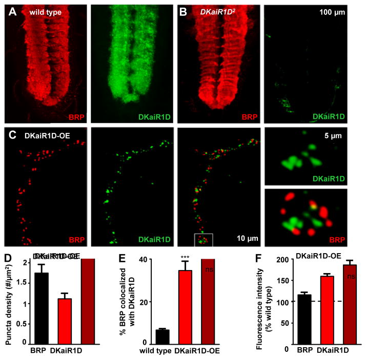
Figure 4. Endogenous DKaiR1D receptors localize to synaptic neuropil and to presynaptic terminals when overexpressed.
Representative images of the ventral nerve cord (VNC) in wild type (A) and DKaiR1D mutants (B) immunostained with anti-DKaiR1D and anti-BRP. DKaiR1D is enriched in synapse-rich areas of the neuropil (highlighted by BRP signal), and is absent in DKaiR1D mutants. (C) DKaiR1D puncta are observed near BRP positive active zones at presynaptic NMJ terminals when overexpressed in motor neurons (DKaiR1D-OE: w;OK6-Gal4/UAS-DKaiR1D) and immunostained with anti-DKaiR1D and anti-BRP. Quantification of BRP and DKaiR1D puncta density in DKaiR1D-OE (D), percent BRP puncta co-localized with DKaiR1D puncta in wild-type synapses and DKaiR1D-OE (E), and fluorescence intensities of BRP and DKaiR1D puncta normalized to wild type backgrounds (F). Error bars indicate ±SEM. Asterisks indicate statistical significance using t-test: (*) p<0.05; (**) p<0.01; (***) p<0.001; (ns) not significant. Detailed statistical information for represented data (mean values, SEM, n, p) is shown in Table S1.

