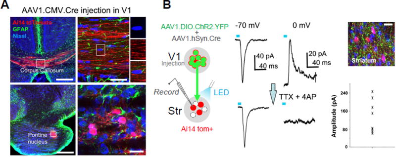
Figure 4. Synaptic specificity of anterograde viral spread.
(A) TdTomato expression (red) and GFAP labeling (green) in the corpus callosum (upper) and pontine nucleus (lower) following injection of AAV1-CMV-Cre into V1 of an Ai14 mouse. High-magnification images (right panels) show that Nissl stained cell bodies (blue) within the corpus callosum were negative for tdTomato (upper), and that tdTomato-positive cell bodies within PN were negative for GFAP staining (lower). Scale bars: 250 µm, left panels; 25 µm, right panels.
(B) Slice recording from transneuronally labeled neurons in the striatum (red) following co-injection of AAV1-hSyn-Cre and AAV1-EF1a-DIO-ChR2-YFP into V1 (left panel). Top right image shows ChR2-expressing axons (green) surrounding tdTomato-labeled striatal neurons. Scale: 25 µm. Middle panel, average LED-evoked excitatory (−70 mV) and inhibitory (0 mV) currents in an example tdTomato+ striatal neuron before and after perfusing in TTX and 4AP. LED stimulation is marked by a blue bar. Right bottom, a summary of amplitudes of average monosynaptic excitatory currents evoked by LED in 9 recorded striatal cells.

