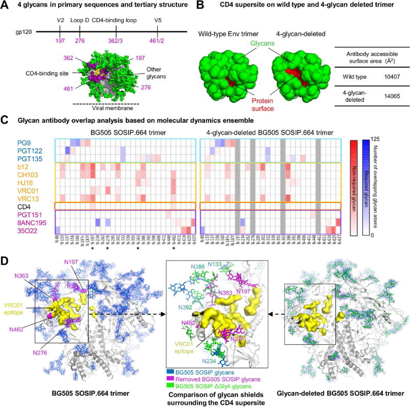
Figure 1. Identification of glycans for targeted removal, with molecular dynamics and crystal structure of a 4-glycan-deleted HIV-1 Env trimer.
(A) Location of 4 glycans (purple) proximal to the HIV-1 CD4-binding site (yellow) in primary sequences and tertiary structure.
(B) Antibody accessible- protein surface area on wild type and glycan-deleted BG505 SOSIP based on molecular dynamics simulation of BG505 SOSIP with Man5 glycans. The antibody accessible-protein surface area was calculated using a 10Å probe with Naccess. Protein and glycan surfaces are shown red and green, respectively.
(C) Glycan-antibody overlap analysis derived from a 500 ns molecular dynamics simulations of Man-5- and 4-glycan-deleted models of the HIV-1 glycan shield. The extent of steric overlap with antibody is shown in blue for those glycans that are known to be required for recognition and in red for glycans not known to be required for recognition.
(D) Comparison of crystal structures BG505 SOSIP (left) and 4-glycan-deleted BG505 SOSIP (right) trimer indicates selective removal of 4 glycans (purple) around the VRC01-binding site (yellow) reduces glycan shielding. Center inset shows the superposition of glycan shield around the VRC01-binding site in wild type and 4-glycan-deleted BG505 SOSIP trimer. Ordered glycan residues are shown in stick representation with 2Fo-Fc electron density for glycans shown at 0.8 σ on gp140 trimer glycans and colored in blue and green for wild type and glycan-deleted BG505 SOSIP trimer, respectively.
See also Figure S1 and Table S1.

