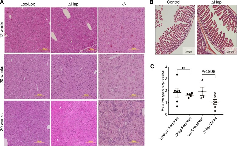
Fig. 3.
Atp7bΔHep mice do not show pathology up to 30 wk, have normal intestinal morphology, and do not upregulate TLR4. A: representative histology (hematoxylin and eosin staining) of liver sections from Atp7bΔHep, Atp7b−/−, and Atp7bLox/Lox mice at different ages illustrates morphologic changes in Atp7b−/− liver, whereas histomorphology for Atp7bΔHep is similar to that of Atp7bLox/Lox mice. B: histology of intestinal sections from 52-wk-old Atp7bΔHep mice and controls demonstrates the lack of pathologic changes in intestine. C: TLR4 mRNA expression in 20-wk liver of Atp7bΔHep, and Atp7bLox/Lox mice (n = 4–7). Data were analyzed using a two-tailed unpaired t-test.

