Fig. 4.
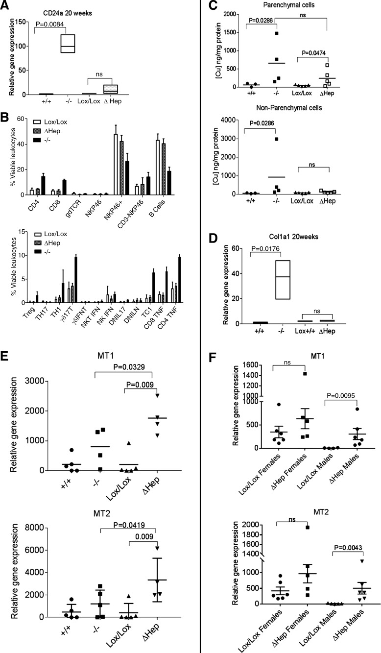
Atp7bΔHep and Atp7b−/− mice differ in copper accumulation in nonparenchymal cells, metallothionein, expression, and immune response. A: expression of CD24a is significantly increased in Atp7b−/−, but not in Atp7bΔHep livers at 20 wk after birth (n = 4–5 per genotype). The qPCR analysis was done using GAPDH as a control, and the amounts of transcripts were normalized to the transcript levels in Atp7b+/+ liver taken as 1. Data were analyzed using unpaired parametric two-tailed t-test. B: profiles of inflammatory cells in livers of 20-wk-old mice Atp7bLox/Lox, Atp7b−/−, and Atp7bΔHep characterized by flow cytometry (n = 3–4 per genotype; Treg, regulatory T cells; IFNT, interferon-τ; NKT, natural killer T cells; DNIL17, double-negative IL-17 and IL-N-producing T cells, respectively; TC1, type 1 CD8+ T cells). C: copper measurements in isolated hepatocytes and nonparenchymal cells. Parenchymal and nonparenchymal cells were isolated from 6-wk-old livers of Atp7b+/+, Atp7b−/−, Atp7bLox/Lox, and Atp7bΔHep mice (n = 3–5 per genotype; data were analyzed using unpaired 1-tailed t-test, because copper levels in Atp7b−/− samples were uniformly higher compared with controls). D: Col1a1 is significantly upregulated in Atp7b−/−, but not in Atp7bΔHep livers at 20 wk. E: differential expression of metallothionein 1 and 2 (MT1 and MT2) in 20-wk Atp7b−/− and Atp7bΔHep livers (n = 4–5). F: expression of MT1 and MT2 in the liver of 20-wk-old Atp7bΔHep males and females and age-matched controls. Data were analyzed using unpaired two-tailed t-test.

