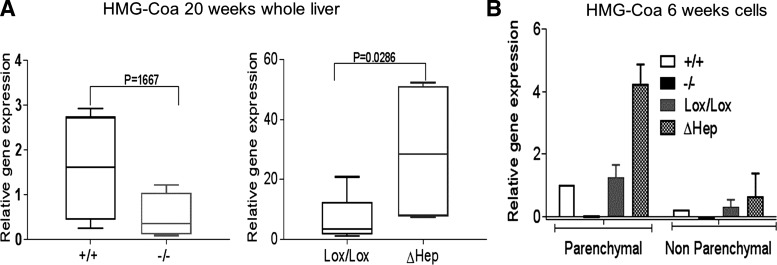
Fig. 6.
Levels of HMG-CoA differ significantly between the Atp7bΔHep and Atp7b−/− strains. A: expression of HMG-CoA reductase in whole livers at 20 wk (n = 4 or 5 per each condition). Gene expression was normalized to Atp7b+/+ levels, and data were analyzed using Mann-Whitney two-tailed t-test. B: expression of HMG-CoA reductase in parenchymal and nonparenchymal cells isolated from livers of 6-wk-old mice (n = 3–5 each for Atp7bLox/Lox and Atp7bΔHep; cells from individual Atp7b+/+ and Atp7b−/− livers are used for reference). The values were normalized to GAPDH and compared with 6-wk-old Atp7b+/+ hepatocytes.

