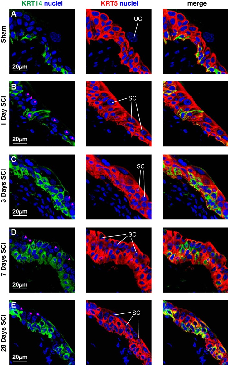
Fig. 8.
Distribution of KRT14 and KRT5 in the urothelium post SCI. Bladders from sham-treated mice (A) or those with SCI (B–E) were fixed and processed for confocal microscopy. Tissues were labeled with antibodies to KRT14 and KRT5, and with TO-PRO-3. A representative region of urothelium was imaged, and a projection of a Z-stack is shown for each condition. Examples of superficial umbrella cells (UC) and small superficial cells (SC) are indicated. Intermediate cells that are KRT14−, but KRT5+ are indicated by solid magenta triangles. The magenta-colored asterisk in B marks a KRT14+ basal cell that is extending a cell projection that terminates at the bladder lumen. The sham and SCI experiments included 3 animals per group and were performed on 5 separate occasions. Representative images are presented.

