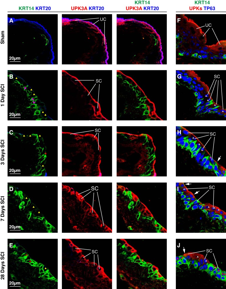
Fig. 9.
Relationship of KRT14, TP63, and UPK expression in small superficial cells post SCI. Bladders from sham-treated mice (A and F) or those with SCI (B–E, G–J) were fixed and processed for confocal microscopy. Tissues were labeled with antibodies to KRT14, KRT20, and UPK3A (A–E), or with antibodies to KRT14, TP63, and UPKs (F–J). A representative region of urothelium was imaged, and a projection of a Z-stack is shown for each condition. Examples of superficial umbrella cells (UC) and small superficial cells (SC) are indicated. Small superficial cells with limited or no KRT14 staining (KRT14−), but positive for UPK3A are marked with yellow-colored triangles, small superficial cells that are KRT14+, but UPK− are indicated by red-colored triangles, and small superficial cells that are TP63+ and UPK+, but KRT14− are marked with gray-colored triangles. The magenta-colored asterisks in B and G mark KRT14+ basal cell that are extending cell projections that terminate at the bladder lumen. The sham and SCI experiments included 3 animals per group and were performed on 5 separate occasions. Representative images are presented.

