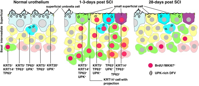
Fig. 12.
Changes in the urothelium in response to SCI. Cellular phenotypes observed in the indicated cell layer of normal (and sham-treated), 1–3 days post SCI, and 28 days post SCI urothelium. Note that the KRT20+ and UPK+ umbrella cells in normal urothelium are replaced in animals with SCI by smaller superficial cells with the indicated phenotypes. At 1 day post SCI in particular, KRT14+ basal cells are observed that send thin projections that terminate in the bladder lumen (one such cell is shown in the middle panel of the figure). This may represent a form of interkinetic nuclear migration. BrdU/MKI67+ nuclei are red-colored and indicate mitotically active cells.

