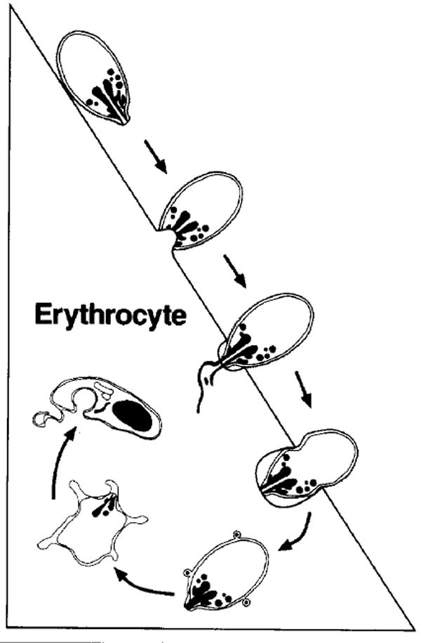
Fig. 3.

Schematic representation of the major morphological events of merozoite invasion. On top, the merozoite is depicted in the attachment and orientation phase, followed by binding, junction formation and rhoptry discharge. The merozoite is then shown entering the parasitophorous vacuole as the junction between the merozoite and the erythrocyte moves towards the posterior end of the merozoite. The parasite is then completely intracellular and flattens with its cytoplasm remaining thick only at the edges, giving the appearance of a ‘ring’.
