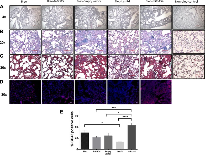
Fig. 8.
miRNA modified B-MSCs and fibrosis in the lung. Mice were injected intratracheally at day 0 with bleomycin or sterile saline (control group, n = 3). At day 7, mice were injected intravenously with control B-MSCs (n = 6) and modified B-MSCs [B-MSCs transduced with an empty vector (n = 6), let-7d (n = 7), and miR-154 (n = 7)] or not received any injection (bleomycin group, n = 6). Mice were euthanized at day 14. Lungs from all the groups of mice were used for histological evaluation using hematoxylin and eosin (A and B) and Masson’s trichrome staining (C). CD45-positive cells (red) over total cells (blue) were assessed in lung slides obtained from the different mice model groups (D and E). Scale bars: 500 µm in A, 100 µm in B and C, 50 µm in D. *P < 0.05. ***P < 0.001. ****P < 0.0001.

