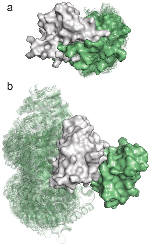
Figure 1.
Docking results for biological and crystallographic dimers. (a) Docking of E. coli met repressor (PDB ID 1cmb, solid surface in grey) to itself. The 100 lowest energy poses (transparent cartoons in green) closely match the actual position of the second subunit, shown as surface in green. Met repressor is a homodimer in solution. (b) Docking of soybean leghemoglobin A (PDB ID 1bin, grey surface) to itself. No low energy docked pose overlaps with the X-ray structure of the second subunit in the dimer (green surface), indicating that there is no stable dimer in solution. Accordingly, soybean leghemoglobin A is a monomer.

