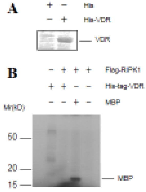
Figure 2. RIPK1 did not phosphorylate VDR in in vitro immunocomplex kinase assays.

A. His-tagged VDR proteins were expressed in bacteria, purified using nickel beads, and stained with Coomassie blue. B. Flag-RIPK1 was transfected into 293T cells and immunoprecipitated with anti-Flag antibodies. In vitro immunocomplex kinase assays were performed with purified His-VDR protein as a substrate. Myelin basic protein (MBP) was included as a positive control.
