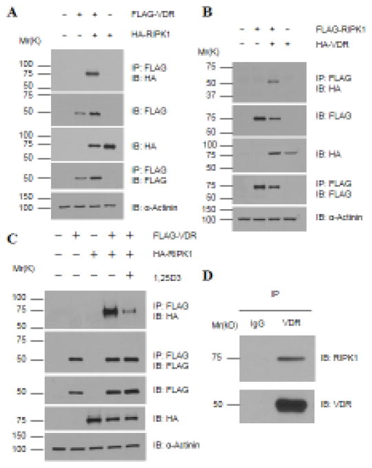
Figure 3. RIPK1 forms a protein complex with VDR, which is decreased by 1,25D3 treatments.
A – C. 293T cells were transfected with 1.5 μg of tagged VDR and RIPK1 as indicated and treated with (Panel C) EtOH or 10−7 M 1,25D3 for 24 hours. Cellular lysates were immunoprecipitated with anti-Flag antibody conjugated beads. Western blot analyses were performed with indicated antibodies. D. Whole cell lysates of L929 cells were immunoprecipitated with anti-VDR antibody followed by Western blot analyses with anti-VDR and RIPK1 antibodies as indicated.

