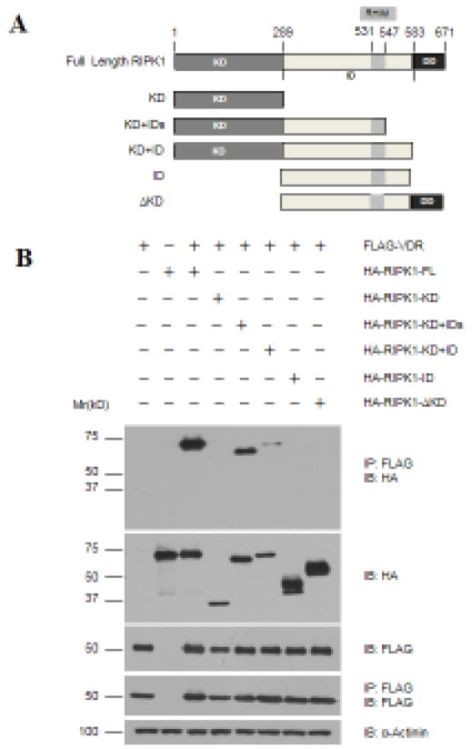
Figure 5. Multiple regions of RIPK1 mediate its binding to VDR.
A. Schematic representation of the domain structure of RIPK1 and its deletion constructs. Amino acid residue numbers are shown at the top of the graphs. KD: kinase domain (aa.1-289); ID: Intermediate domain (aa.290-583); IDs: Smaller ID (aa.290-547); ΔKD: Kinase deleted (aa.290-583). B. 293T cells were transfected with 1.5 μg of Flag-VDR together with 1.5 μg of full-length (FL) or deletion constructs of HA-RIPK1 as indicated. Cellular extracts were subjected to co-immunoprecipitation analyses with antibodies as indicated.

