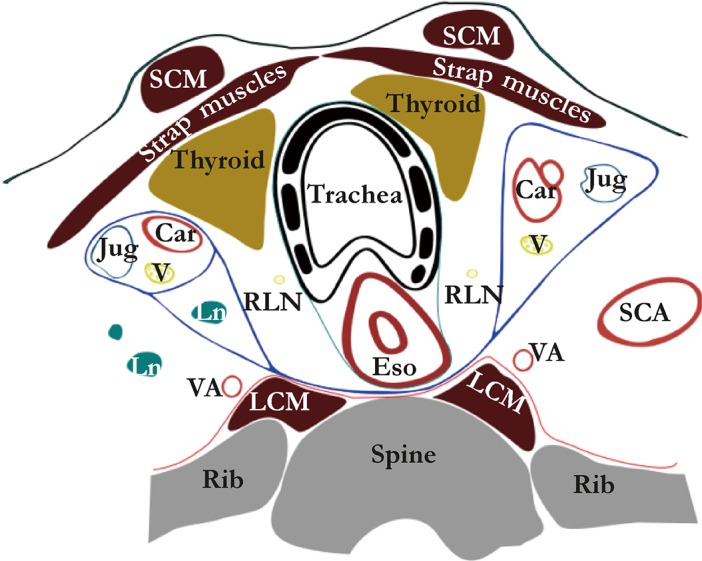
Figure 2.

Peri-esophageal connective tissue layers above the aortic arch (Weijs et al. 2016, rights obtained). The blue line represents the alar fascia and the carotid sheaths; the green line represents the visceral fascia and the red line represents the perivertebral fascia. Car, carotid artery; Eso, esophagus; Jug, internal jugular vein; LCM, longus colli muscle; Ln, lymph node; Rln, recurrent laryngeal nerve; SCA, subclavian artery; SCM, sternocleidomastoid muscle; V, vagus nerve; VA, vertebral artery.
