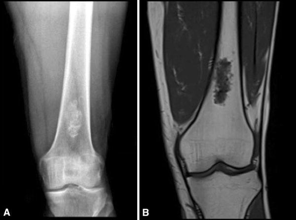
Fig. 1A–B.

A 39-year-old man without any previous pain obtained plain radiographs after a knee contusion while playing soccer. He had no pain at the distal femur or any other relevant findings on physical examination. (A) An AP radiograph of his distal femur shows a central lesion with a cartilaginous matrix and calcifications. (B) A coronal T1 MR image shows a 2 × 4-cm cartilaginous tumor without any cortical compromise or soft tissue mass. This was interpreted as benign by all evaluators.
