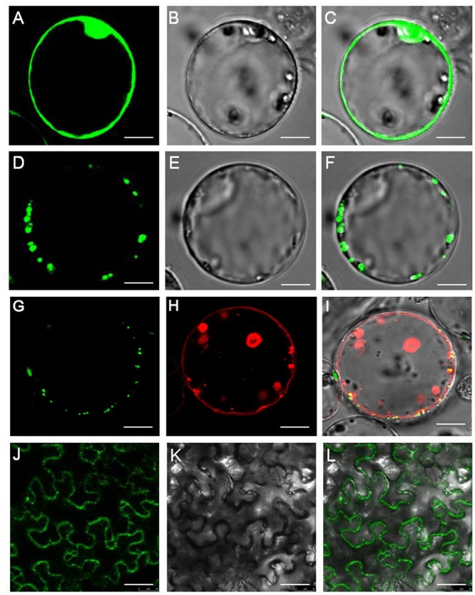
FIGURE 1.

OsNPF7.3 is located in the rice vacuolar membrane. (A–C) Free GFP expression in rice protoplasts. (D–F) OsNPF7.3-GFP expression in rice protoplasts. (G–I) Fluorescence of OsNPF7.3-GFP coexpressed with the vacuolar membrane marker AtTPK-mkate in transiently transformed rice protoplasts. (J–L) Fluorescence of OsNPF7.3-GFP expressed in tobacco epidermal cells. Presented here are fluorescence images of GFP (A,D,G,J), corresponding bright-field images (B,E,K), AtTPK-mkate fluorescence images (H) and merged images (C,F,I,L). Bars = 5 μm for (A–L).
