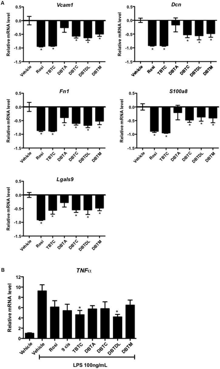
FIGURE 4.
Dibutyltins inhibited the expression of inflammatory genes. (A) 3T3-L1 cells were differentiated with insulin, and exposed to vehicle (DMSO), Rosi 0.1 μM, TBTC 0.1 μM, DBTA 0.1 μM, DBTC 0.1 μM, DBTDL 1 μM, or DBTM 0.1 μM. After 14 days, the cells were collected for gene expression evaluation of Vcam1, Dcn, Fn1, S100a8, and Lgals9 by real-time quantitative PCR. Data are expressed as fold activation relative to transcript levels in vehicle samples (DMSO). ∗p ≤ 0.01 (compared to vehicle samples). (B) Raw 264.7 Cells were exposed to vehicle (DMSO), Rosi 10 μM, 9-cis-retinoic acid (9-cis) 10 μM, TBTC 0.1 μM, DBTA 1 μM, DBTC 0.1 μM, DBTDL 1 μM, or DBTM 0.1 μM. After 4 h, LPS 100 ng/mL was added and maintained for an additional 24 h. After that, the cells were collected for evaluation of gene expression of TNFα by real-time quantitative PCR. Data are presented as mean (SD) of three independent experiments conducted in triplicate and expressed as activation relative to transcript levels in vehicle samples (DMSO). ∗p ≤ 0.01 (compared to vehicle + LPS samples).

