
Figure 3.
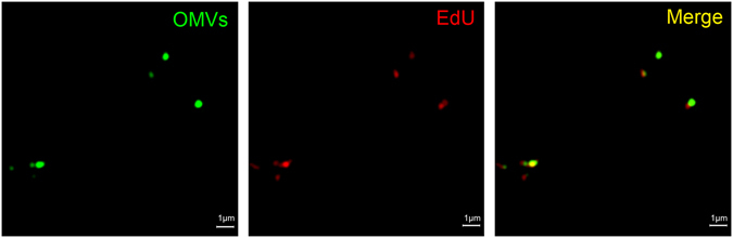
Super resolution microscopy of OMVs carrying bacterial DNA. OMVs were labeled with the lipophilic stain, DiO (green), which targets the OMV membrane, while EdU-incorporated DNA (red) was labeled by a 2-step process. The first step involved incubation with 12.5 μM biotin azide, during which the azide moiety was bound specifically to the alkyne backbone of the EdU molecule in the presence of a copper catalyst. The second step involved incubation with 5 μg/ml streptavidin-conjugated 568 Alexa Fluor® to fluorescently label the biotin-azide bound to the EdU. Scale bar = 1 μm.