
Figure 5.
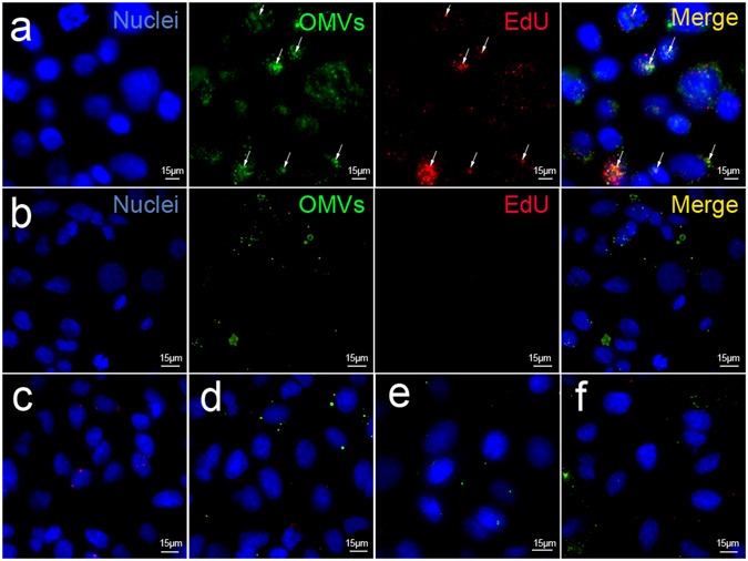
Confocal microscopy showing internalization of OMV-associated DNA in A549 lung epithelial cells. EdU-labeled OMVs, either (a) untreated or (b) DNase-treated, were labeled with the lipophilic stain DiO then added to A549 cells for 5 h, washed to remove non-internalized OMVs, fixed and permeabilized. EdU was detected in a 2-step reaction consisting of incubation with biotin azide, in which the azide binds specifically to the alkyne backbone of EdU, followed by fluorescent detection with streptavidin-conjugated Alexa-Fluor 568. Co-localization of DiO (green) and EdU (red) labels in internalized OMVs are indicated by arrows. Nuclei are stained with DAPI (blue). Merged images showing control samples in which either: (c) biotin azide and streptavidin-AlexaFluor 568 only, (d) no biotin azide or (e) DiO only were added to cells. (f) Non-permeabilized cells. Scale bar = 15 μm.