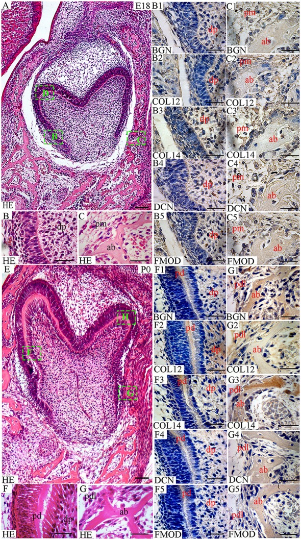
Figure 1.

Localization of FACITs and SLRPs in the developing tooth and surrounding structures at embryonic day 18 (E18) and perinatal day (P0). (A) Haematoxylin and eosin staining of embryonic tooth and surrounding tissues (E18). Details from boxes (B–D) in A is shown in B,C, and Figure 2D. (B) Detail of dental pulp. (C) Detail of peridental mesenchyme and alveolar bone. Expression of biglycan in dental (B1) and peridental mesenchymes (C1). Expression of collagen XIIa in dental (B2) and peridental mesenchymes (C2). Expression of collagen XIVa in dental (B3) and peridental mesenchymes (C3). Expression of decorin in dental (B4) and peridental mesenchymes (C4). Expression of fibromodulin in dental (B5) and peridental mesenchymes (C5). (E) Haematoxylin and eosin staining of the tooth and surrounding tissues at P0. Details from boxes (F–H) in E is shown in F,G, and Figure 2H. (F) Detail of dental pulp and predentin. (G) Detail of periodontal ligaments and alveolar bone. Expression of biglycan (F1), collagen XIIa (F2), collagen XIVa (F3), decorin (F4), and fibromodulin (F5) in predentin and dental pulp. Expression of biglycan (G1), collagen XIIa (G2), collagen XIVa (G3), decorin (G4), and fibromodulin (G5) in the periodontal ligament and alveolar bone. Dp, dental pulp; pm, peridental mesenchyme; ab, alveolar bone; pd, predentin; pdl, periodontal ligaments; BGN, biglycan; COL12, collagen type XIIa; COL14, collagen type XIVa; DCN, decorin; FMOD; fibromodulin; HE, haematoxylin/eosin. Scale bar (A,E) = 100 μm. Scale bar (B,B1–B5,C,C1–C5,F,F1–F5,G,G1–G5) = 25 μm.
