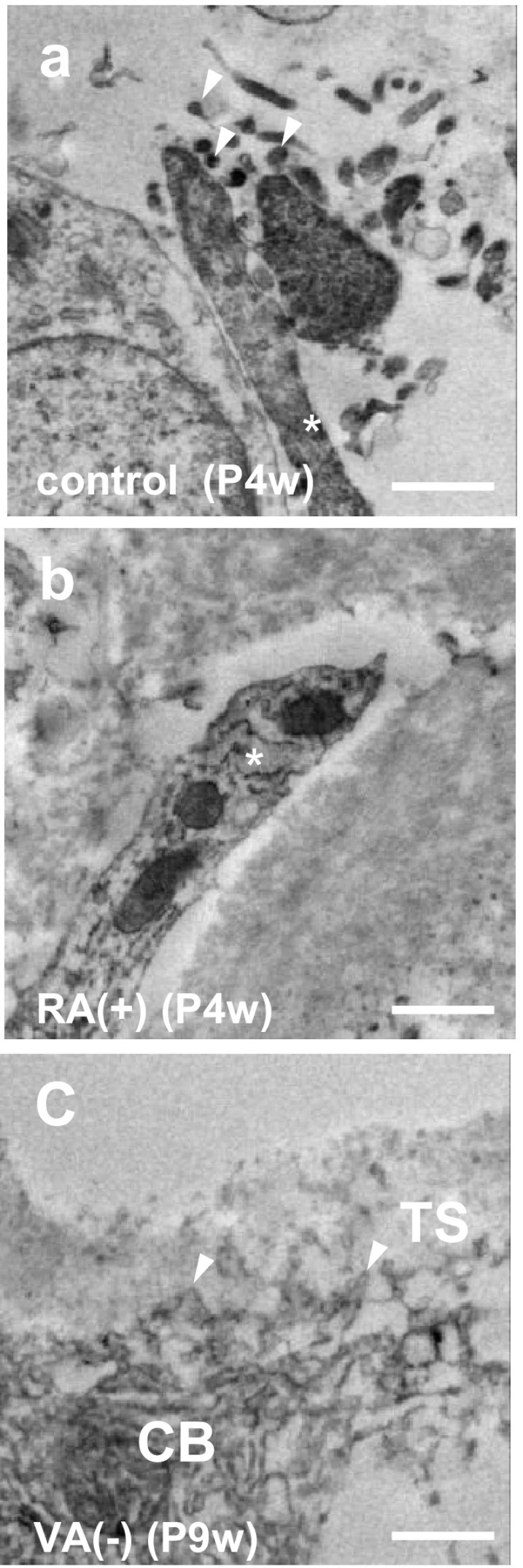
Fig. 5.

Process apex of septoclasts. Immunoelectron micrographs of E-FABP-immunoreactive septoclasts at the COJ of the GP of mouse proximal tibiae of P4w control (a), P4w RA excess (b), and P9w VA deficiency (c). Asterisks septoclastic processes; arrowheads: microvilli (a) or short spines of septoclastic cell body (c); CB septoclastic cell body, TS transverse septum; scale bars 1 μm
