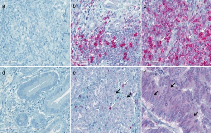
Fig. 1.
Programmed cell death protein 1 (PD1) expression on tumor-infiltrating lymphocytes (TILs) (a–c) and tumor cells (d–f) in esophageal adenocarcinoma detected by immunohistochemistry. a Negative PD1 staining of TILs. b 2+ (26–50%) positive staining of TILs. c 3+ (51–75%) positive staining of TILs. d Negative staining of tumor cells. e 2+ (26–50%) positive staining of tumor cells. f 4+ (75–100%) positive staining of tumor cells. The immunoreactivity for PD1 of tumor cells and TILs was examined at ×400 magnification, and the staining rate (percentage of tumor cells and lymphocytes showing positive staining, 0–100%) was determined. Arrows indicate examples for a positive PD1 staining on TILs (white arrow) and tumor cells (black arrow)

