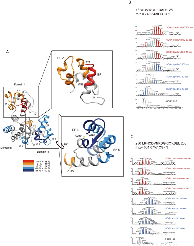
Figure 2.

Structural changes in apo and Ca2+-bound hSCGN analyzed by hydrogen/deuterium exchange mass spectrometry. (A) Overlay of differential HDX data onto the structures of human SCGN structure which were modeled in silico with the MODELLER 9.9 program. Percentage difference in HDX between apo and Ca2+ bound hSCGN is colored according to the key. The differential deuterium exchange patterns of hSCGN peptide sequences representing EF-hand and Cys residue in N-terminal (B) and C-terminal (C) peptides of hSCGN.
