
Figure 1.
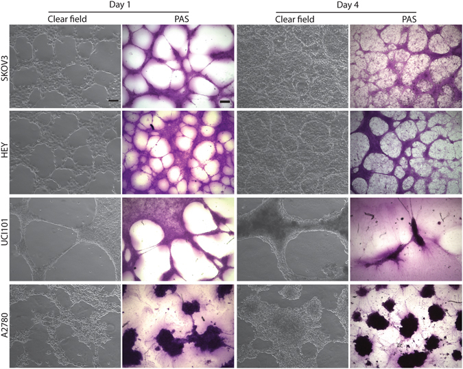
Ovarian cancer cell lines form tubular structures in 3D culture. Clear field images (magnification: 10×. Scale bar: 100 μm) and PAS stained images (magnification: 4×. Scale bar: 200 μm) of 3D cultures on matrigel at day 1 (left panels) and day 4 (right panels) in SKOV3, HEY, UCI101 and A2780 cells. At day 1 all cell lines form structures that could be described as capillary-like and are PAS+, however at day 4 only SKOV3 and HEY cell lines form cell aggregates surrounded by capillary-like structures. Tubular structures in SKOV3 and HEY were more PAS+ compared to the monolayer of cells, however in UCI101 and A2780, the individual cells stained strongly for PAS.