
Figure 3.
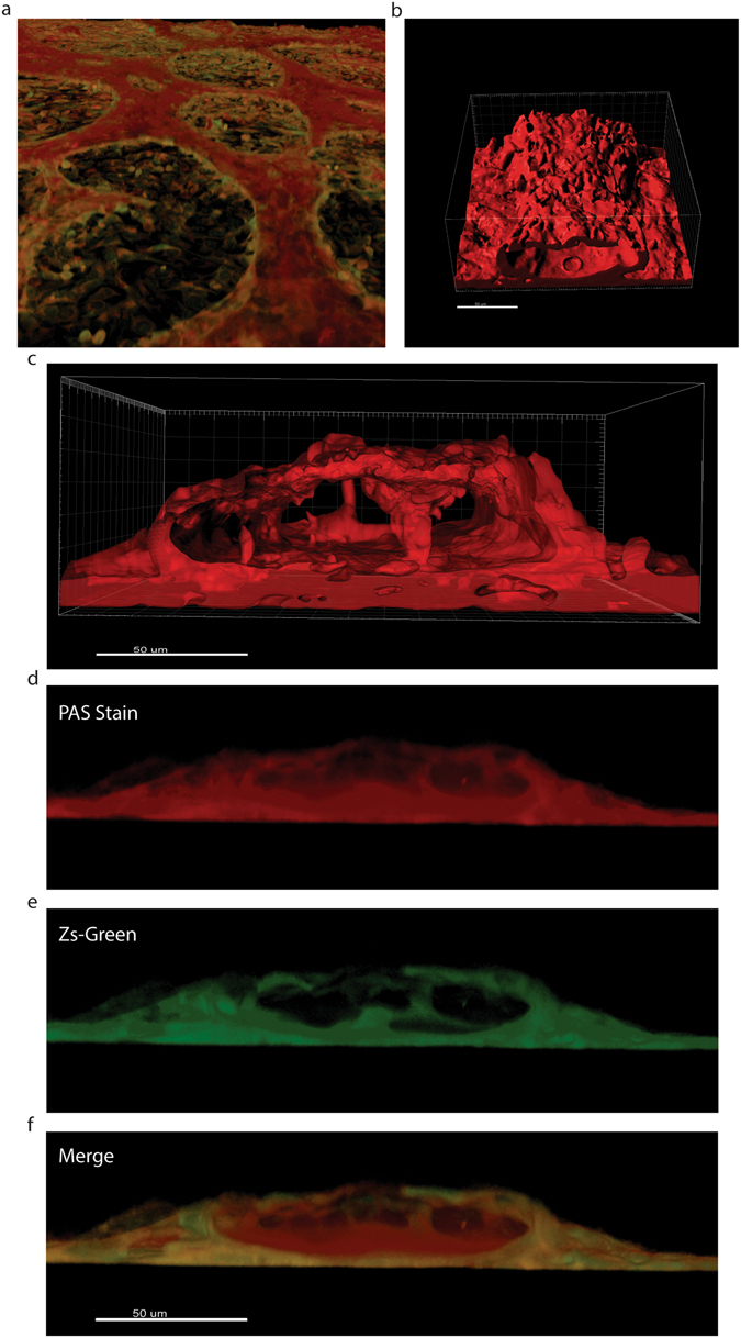
Tubular Structures possess a glycoprotein-rich internal layer. (a) 3D reconstruction of the monolayer and tubular structures formed by the HEY-GFP ovarian cancer line upon PAS staining and analysis with laser confocal microscopy. PAS emits within the red spectrum upon laser excitation at 560 nm. (b) Reconstruction of the glyoprotein-rich component of a tubular structure. (c) Reconstruction of the glyoprotein-rich component surrounding a lumen. (d,e) Cross-section of PAS stained tubular structures. In red (panel d) a glycoprotein-rich component constructed from PAS staining. In green (panel e) cancer cells forming a tubular structure, (f) merge of the previous panels showing that the glycoprotein-rich component is present on luminal side of the green (cancer cell) tubular structures.