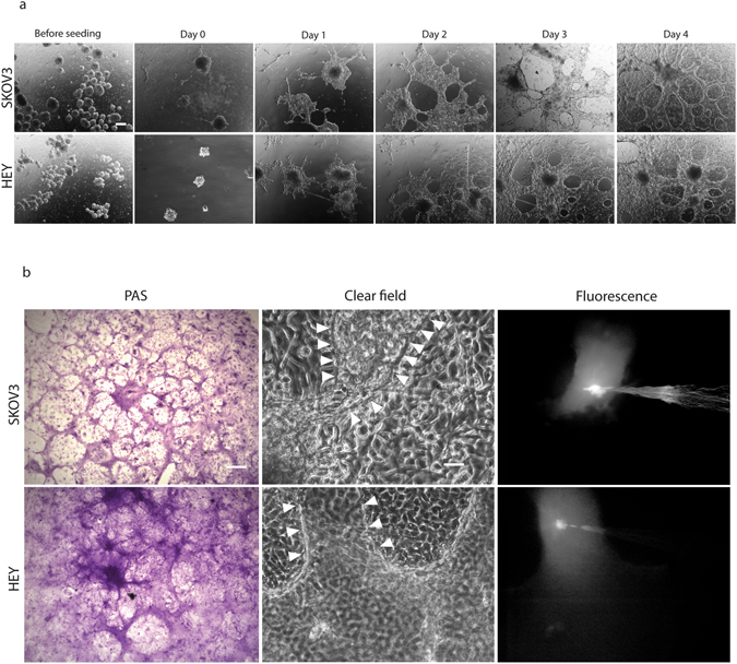
Figure 6.

Tubular structure formation in ovarian cancer cell-derived spheroids. (a) Cancer spheres were generated from parental SKOV3 and HEY cells (noted as “before seeding”) and individual spheres seeded onto matrigel. At day 1 the cells grew principally within the spheres structure before spreading out to cover the dish at day 3 and forming clear tubular structures at day 4 in both cell lines. Magnification: 4×. Scalebar: 200 μm. (b) PAS staining (left) and microinjection (middle and right) of a 4 day-old spheres initiated 3D-cultures on matrigel. Arrowheads line the borders of microinjected structure. Magnification: 10×. Scalebar: 100 μm.
