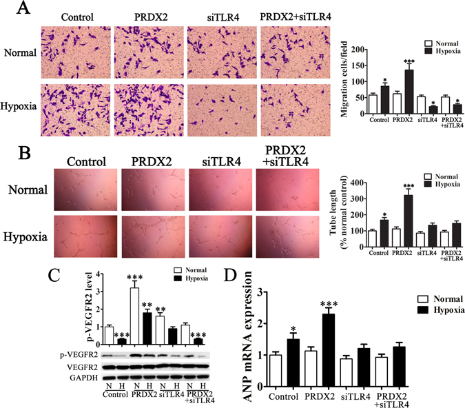
Figure 4.

PRDX2 overexpression in H9c2 cells promote HUVEC migration, vasculogenic mimicry formation and myocardial hypertrophy related protein expression. H9c2 cells were cultured in serum-free medium and exposed to hypoxic conditions for 6 h then in normal conditions for another 24 h, then the medium was harvested for transwell and vasculogenic mimicry formation of HUVECs. (A) Migration of HUVECs was assessed using transwell chambers. The invasive capabilities of HUVECs are significantly increased with chemotaxis of PRDX2 overexpression in H9c2 cells. The data are presented as means ± SD of the mean from ten separate cell experiments. *p < 0.05, **p < 0.01, ***p < 0.001 vs control. (B) Images of tube formation. Tube length is presented as percent of total tube length per field versus untreated control cells. The data are presented as means ± SD of the mean from ten separate cell experiments. *p < 0.05, **p < 0.01, ***p < 0.001 vs control. (C) VEGFR2 phosphorylation in the surface of HUVECs was detected by western blots. The phospho-specific bands were quantified and normalized by the intensities of the corresponding VEGFR2 bands. The data are presented as means ± SD of the mean from three separate cell experiments. *p < 0.05, **p < 0.01, ***p < 0.001 vs control. (D) RT-PCR analysis of ANP expression. The data are presented as means ± SD from three separate cell experiments. *p < 0.05, **p < 0.01, ***p < 0.001 vs control.
