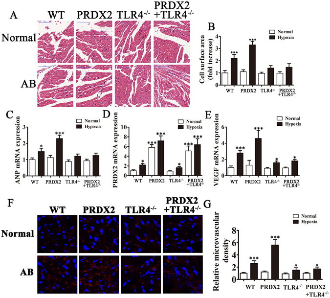
Figure 6.

The overexpression of PRDX2 promote angiogenesis and myocardial hypertrophy. After 2 weeks of coronary artery ligation, serum from mice was harvested for RT-PCR analysis and myocardial tissue were harvested 4 weeks after artery ligation for immunohistochemical analysis. (A) Effects of PRDX2 on cell surface area of cardiomyocytes. (B) Cell surface area was calculated by measuring 10 random cardiomyocytes. (C–E) RT-PCR for ANP (C), PRDX2 (D) and VEGF (E) expression. The data are presented as means ± SD from three separate experiments from five to six mice in each group. *p < 0.05, ***p < 0.001 vs normal WT group. (F) The formation of microvascular cells in infarction region were stained with CD31. AB, aortic banding. The data are presented as means ± SD from 10 fields in each group. *p < 0.05, ***p < 0.001 vs normal WT group.
