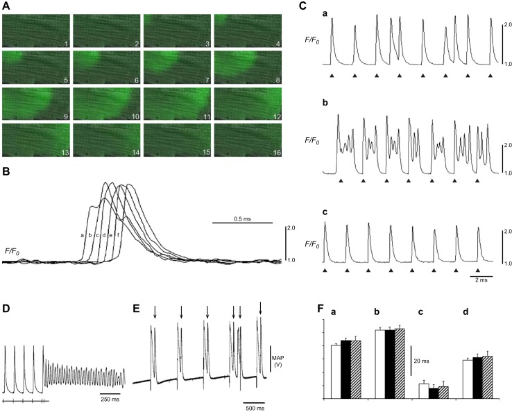
Figure 15.
Epac-induced RyR2 activation as an exemplar for Ca2+-mediated arrhythmia. A: propagation of a Ca2+ wave from one end of the cell to the other following 8-CPT challenge shown in successive confocal microscope frame scan images. B: Ca2+ fluorescence signal from six successive 2 μm × 2 μm regions of interest (a–f), placed along the long axis of a myocyte demonstrating progressively increasing delays in onset of the Ca2+ transient. C: Ca2+ signals in regularly stimulated ventricular myocytes (triangles mark timing of pacing stimuli) showing irregularly occurring ectopic Ca2+ transients during 8-CPT treatment (a, b) abolished by KN-93 (c). D: persistent ventricular tachycardia (VT) following programmed electrogram stimulation observed during perfusion with 8-CPT prevented by CaMKII inhibition with KN-93. E: triggered activity (*) during intrinsic activity observed during perfusion with 8-CPT, prevented by KN-93 pretreatment. F: epicardial (a) and endocardial APD90 (b), ΔAPD90 (c), and VERP (d) under control conditions (clear bars), during 1 μM 8-CPT treatment in the absence (black bars) and following 1 μM KN-93 pretreatment (striped bars). [From Hothi et al. (446).]

