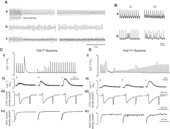
Figure 17.
Physiological features mediating arrhythmogenesis in Pak1-deficient hearts. A: monophasic action potential (AP) recordings showing AP alternans (a), torsades de pointes (TdP) (b), and polymorphic VT (c) following burst pacing (horizontal line below trace) in ex vivo Pak1-cko hearts, features not observed in Pak1-f/f hearts. B: Ca2+ transients in field-stimulated Pak1-f/f (a) and Pak1-cko myocytes (b) at a 1-Hz stimulation frequency under baseline (left traces; i) and chronic β-adrenergic stress conditions (right traces; ii). Increased pacing frequencies increase the occurrence of Ca2+ waves to greater extents in Pak1-cko than Pak1-f/f myocytes particularly with chronic β-adrenergic stress. C and D: recovery of SR Ca2+ stores from after previous depletion by caffeine challenge, after which regular stimulation resumed. i: Ca2+ transients indicating recovery of SR Ca2+ in Pak1-f/f (C) and Pak1-cko myocytes (D) under baseline conditions. a–c: Comparison of increasing Ca2+ transients (ii), constant ICaL (iii), and increasing INCX (iii) at different stages (a–c) of SR Ca2+ recovery. [From Wang et al. (1230).]

