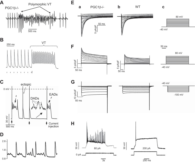
Figure 19.
Arrhythmogenic features of PGC1β−/− hearts. A: increased heart rate following isoproterenol challenge accompanied by polymorphic ventricular tachycardia (VT) in PGC1β−/− mice during ECG recording. B: monophasic action potential (AP) recordings of VT following programmed electrical stimulation in Langendorff-perfused PGC1β−/− hearts. C: APs from PGC1β−/− ventricular myocytes showing early (EADs) and delayed afterdepolarizations (DADs) and ectopic APs. Inset magnifies voltage trace 40-fold. D: abnormal Ca2+ homeostasis with intermittent elevations in diastolic Ca2+ and Ca2+ waves increased in amplitude and frequency in isoproterenol challenged PGC1β−/− ventricular myocytes. E: voltage-gated ICa and transient and sustained outward IK (F) in response to depolarizing pulses from −40 to +50 mV altered in successive 10-mV increments from a −40 mV holding potential. G: inwardly rectifying currents obtained in response to hyperpolarizing steps from −40 to −100 mV incremented in 10-mV intervals. E–G shown for PGC1β−/− (a) and WT ventricular myocytes (b), with voltage step protocols shown in c. H: step current injections produced single APs with prolonged plateaus with burst AP firing in PGC1β−/− (a) but not WT myocytes (b). [From Gurung et al. (387).]

