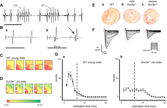
Figure 7.
Conduction and arrhythmic properties in ageing male Scn5a+/− hearts. A: lead II electrocardiographic traces obtained from anesthetized aged Scn5a+/− mouse showing spontaneous nonsustained ventricular tachycardia (VTs indicated by arrow). B: chest lead ECG complexes from young (a) and old (b) intact anesthetized male Scn5a+/− mice. The latter shows patterns of fragmented QRS complexes, indicating bundle branch block most frequently observed with Scn5a+/−. C and D: activation maps from five successive cardiac cycles in young male WT (C) and old male Scn5a+/− hearts (D). E: picrosirius red staining demonstrating ventricular fibrosis in 85-wk-old WT (a) and Scn5a+/− with mild (b) and severe fibrosis which appears red (c). F: corresponding INa records in ventricular myocytes from 12-wk-old WT (a) and mildly (b) and severely affected Scn5a+/− mice (c). G: frequency distributions of activation times in young male WT (a) and old male Scn5a+/− mice (b). [From Jeevaratnam and co-workers (501–503) and Leoni et al. (642).]

