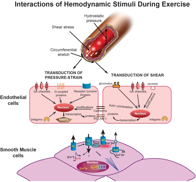
FIGURE 2.
Illustration showing the interactions of hemodynamic signals (top) (hydrostatic pressure, shear stress, and circumferential stretch) that modulate vascular adaptation to exercise. The effects of pressure and/or stretch on the endothelial cells are shown in the middle as described in the text. At the bottom, the figure illustrates exercise-induced adaptations of smooth muscle cells. The middle left of the smooth muscle figure shows calcium transients with decreased intracellular calcium ([Ca2+]i) response to selective agonists (e.g., endothelin) in exercise-trained cells in red (which produces a reduced Ca2+-dependent activation of contraction). This decreased [Ca2+]i occurs despite an increased Ca2+ influx through L-type Ca2+ channels (Cav1.2). Nuclear Ca2+ responses ([Ca2+]n) are also reduced by exercise training, which may affect Ca2+-dependent transcription factors (CaTF; e.g., CREB, NFAT) and target gene expression. Also illustrated is the increased spontaneous, slow-Ca2+ release from the SR into the subscarolemmal space ([Ca2+]ss) which may contribute to the increased activation of large-conductance, Ca2+-activated (BK) potassium channels caused by exercise. Voltage-gated (Kv) potassium channels are also activated by exercise training. [Bottom panel modified from Bowles and Laughlin (32) and Langille and O'Donnell (167), with permission from American Heart Association.]

