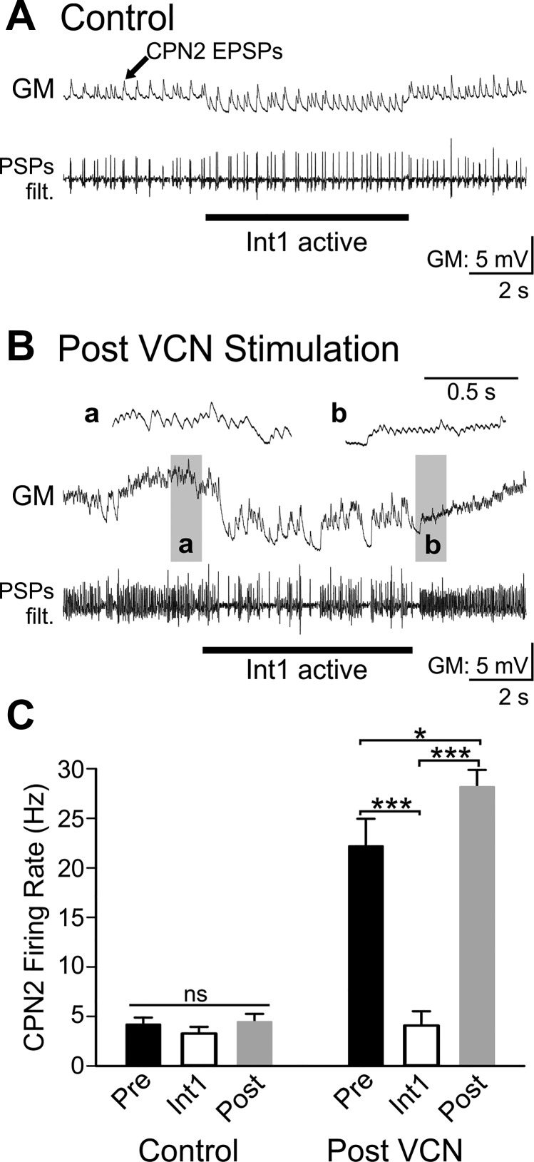
Fig. 8.
Int1 inhibits CPN2 and triggers post inhibitory rebound after VCN stimulation, but not in control conditions. A: in control conditions there was weak CPN2 activity recorded as excitatory postsynaptic potentials (EPSPs) in the GM neuron. A filtered version of the GM recording (PSPs filt.) is shown below the original trace. The Int1 active epoch did not alter the CPN2 firing rate, and there was no postinhibitory rebound (PIR) firing after Int1 activity was terminated. Int1 activity was controlled with depolarizing and hyperpolarizing current injections into LG due to its inhibition of Int1 (see materials and methods). GM is electrically coupled to LG and thus hyperpolarized with LG during the Int1 active period. B: post-VCN stimulation, CPN2 firing rate was higher but was decreased during the Int1 active epoch. After the Int1 active epoch CPN2 fired at a higher rate than before activation of Int1. a And b: gray boxed areas at an expanded time scale. C: the average CPN2 firing rate was not altered by Int1 in control conditions (Control Pre vs. Int1) nor was there a rebound in firing rate elicited after a period of Int1 activity (Control Pre vs. Post, n = 7). VCN stimulation triggered increased CPN2 activity. Under this condition, Int1 inhibited CPN2 activity and triggered a rebound in CPN2 firing rate (n = 6). Black bars: Pre-Int1 activity; white bars: Int1 active; gray bars: Post-Int1 activity during control conditions (left) and after VCN stimulation (right). nsP > 0.05, *P < 0.05, ***P < 0.001.

