Fig. 9.
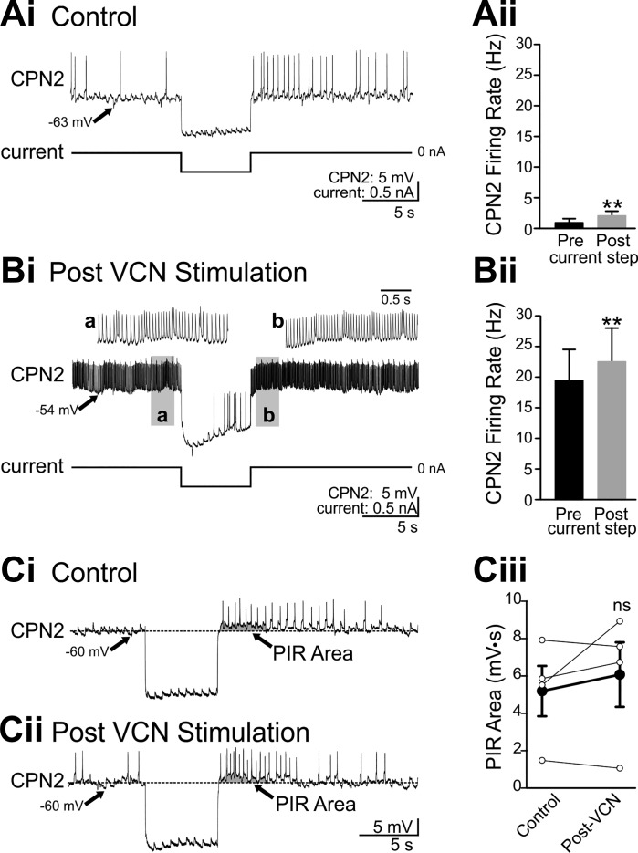
CPN2 expresses similar post inhibitory rebound properties in control conditions and after VCN stimulation. Hyperpolarizing current injection into CPN2 in control conditions elicited rebound firing after the end of the current step in an example recording (Ai) and across preparations (Aii) (n = 6). After VCN stimulation, the same amplitude current step decreased CPN2 firing and triggered rebound firing after the end of the current step in the example recording (Bi) and across preparations (Bii) (n = 5). a And b: gray boxed areas at an expanded time scale. A step current injection elicited rebound firing in CPN2 in a different preparation than above (A and B) in control conditions (Ci) and after VCN stimulation (Cii), when steady current injection was used to maintain CPN2 at the same membrane potential as in control. To compare PIR in control and post-VCN conditions at the same CPN2 membrane potential, “PIR area” (arrow, gray shading) was measured (see materials and methods). Ciii: there was no consistent change in PIR area (n = 4). EPSPs evident in CPN2 in A–C are due to spontaneous action potentials in the AGR sensory neuron. **P < 0.01, nsP > 0.05.

