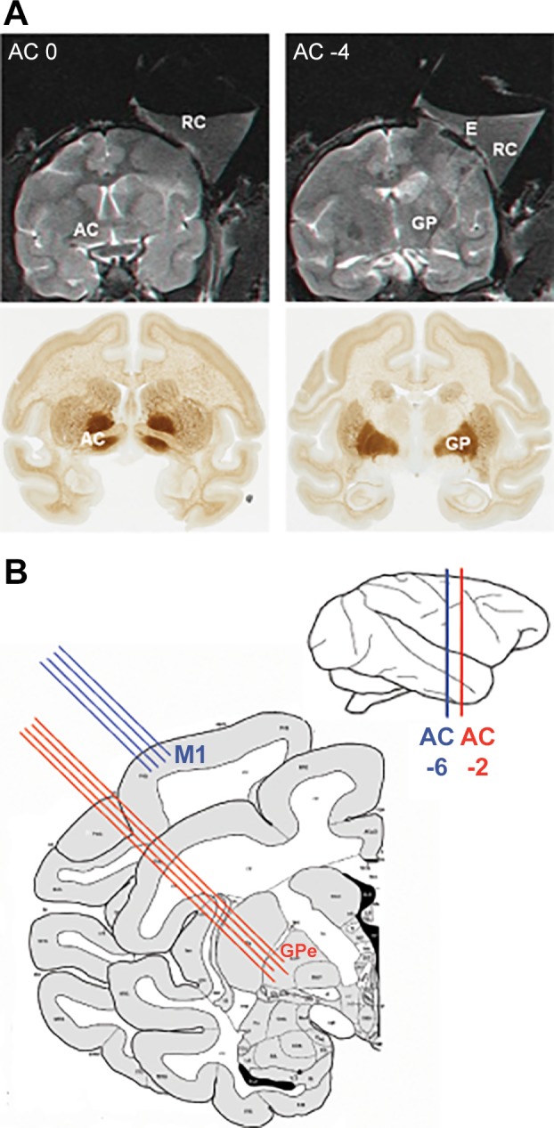
Fig. 1.

Anatomical localization and experimental setup. A: coronal MRI images (top) numbered with respect to distance (in mm) from the anterior commissure. The MRI scan was performed with 5 tungsten electrodes inserted at known coordinates of the recording chamber (2 electrodes can be seen at top right). The coronal MRI images are aligned with coronal sections (bottom) of the African green monkey atlas (www.brainmaps.org). B: experimental setup. Extracellular activity from M1 and the GPe was recorded simultaneously by 4 electrodes in each structure. The atlas scheme is adapted from BrainInfo (1991–present), National Primate Research Center, University of Washington (http://www.braininfo.org). AC, anterior commissure; E, electrode; GP, globus pallidus; GPe, external globus pallidus; M1, primary motor cortex; RC, recording chamber.
