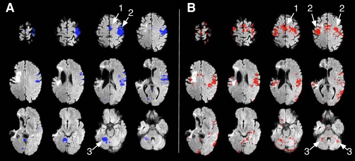
Fig. 1.
Functional magnetic resonance imaging of movements of the unaffected (A) and affected (B) fingers. Areas active during finger movement are thresholded at z = 3.1–12 and superimposed on a T2-weighted structural FLAIR image showing the infarction in the distribution of the right middle cerebral artery. Movement of the unaffected fingers of the right arm (A) is associated with neural activation in the contralateral motor area (1), contralateral sensorimotor cortex (2), and the contralateral supplementary motor cortex and cerebellar hemisphere (3). Movement of the affected fingers of the left hand (B) is associated with widely distributed neural activation in both supplementary motor areas (Smith et al. 2004), sensorimotor cortex (Jenkinson et al. 2002) and cerebellar hemispheres (Smith 2002). The brain images are shown in radiological convention (left side of the image shows right hemisphere).

