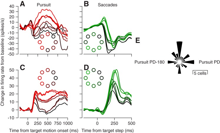
Fig. 3.
Comparison of direction tuning in oculomotor vermis for pursuit and saccadic eye movements. A and C: simple-spike firing vs. time during pursuit in 8 directions for 2 example Purkinje cells. B and D: simple-spike firing vs. time during 10° amplitude saccades in 8 directions for the same 2 Purkinje cells. Colors of traces correspond to colors of dots in each inset and indicate the direction of the target motion for pursuit or the target step for saccades. E: polar histogram indicating the distribution of the difference in preferred direction (PD) of vermis Purkinje cells for saccades vs. pursuit.

