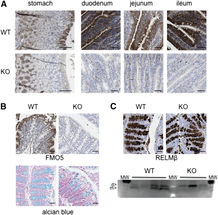
Fig. 4.
Expression of FMO5 and RELMβ in mouse intestine. (A) Immunohistochemical analysis of sections of stomach and small intestine of 30-week-old mice. Sections were incubated with an antibody against FMO5. Top panels, WT mice; bottom panels, Fmo5−/− (KO) mice. Scale bar, 100 μm. (B) Immunohistochemical analysis of sections of colons from 30-week-old mice. (Top panels) Sections incubated with antibodies against FMO5. (Bottom panels) Alcian blue staining of sections of colons from 30-week-old mice. Left-hand panels, WT mice; right-hand panels, KO mice. Scale bar, 100 μm. (C) Immunohistochemical analysis of sections of colons of 30-week-old mice. (Top panels) Sections incubated with antibodies against RELMβ. (Bottom panel) Western blot analysis of colonic content proteins from four WT and three KO mice. d, dimer; m, monomer; MW, molecular weight standards. Protein loading was assessed, and the blot was developed as described in Materials and Methods.

