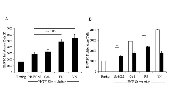
Figure 5.

HGF induced HMVEC proliferation requires the Map kinase pathway. Panel A-HMVEC in MCDB-131 medium was plated on poly-D-lysine coated 48-well plates at a density of 2.5 × 10³ cells/well and were stimulated with HGF (10 ng/ml) in the presence and absence of ECM molecules FN, VN or collagen-1. Basal proliferation was measured with cells treated with non-supplemented medium. Cell numbers were quantified 48 hours post-stimulation using CyQuant reagent. Data is presented as cell numbers with the increase of proliferation of HGF-FN and HGF-VN treated samples significant compared with cells treated with HGF alone as determined by a one-way ANOVA (p < 0.05) n = 3. Panel B-HMVEC were plated onto poly-D-lysine coated wells (1 × 10⁴/well) overnight in supplemented medium. Cells were then incubated with medium comprising 0.1% FBS plus HGF (20 ng/ml) in the presence or absence of ECM proteins (10 μg/ml) as shown containing no inhibitor (white bars) or the MEK inhibitor U1026 (10 μM, black bars). Cells numbers were quantified after a further 48 h using CyQuant reagent.
