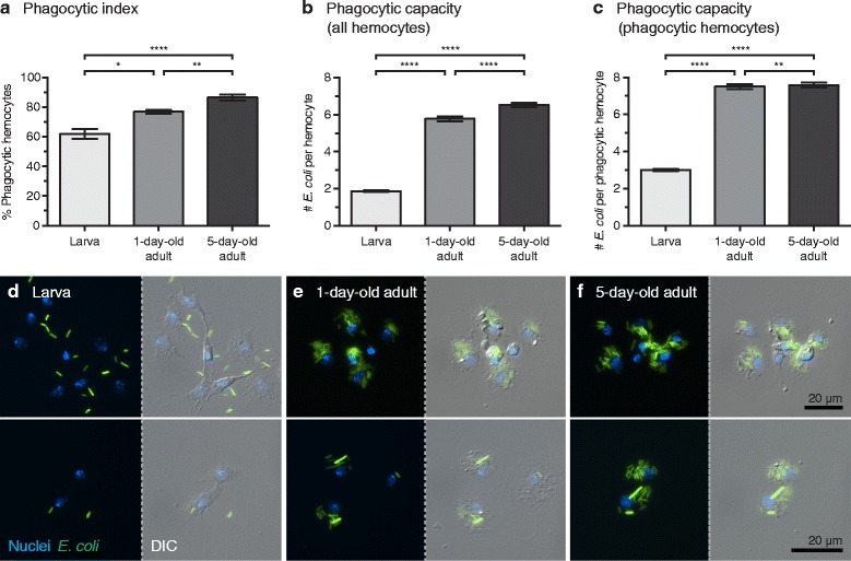
Fig. 4.

Hemocyte phagocytic burdens and spread in adults and larvae. a Percentage of larval and adult hemocytes that phagocytosed bacteria at 1 h post-infection with E. coli (phagocytic index). b, c Number of bacteria in individual larval or adult hemocytes at 1 h post-infection (phagocytic capacity). Data were analyzed for all hemocytes observed (b), and for only the hemocytes that had engaged in phagocytosis (c). Data were analyzed by the Kruskal-Wallis test, followed by Dunn’s post-hoc test (*P < 0.05, **P < 0.01, ****P < 0.0001). In a-c whiskers denote the SEM. d-f Larval and adult hemocytes viewed under both fluorescence and DIC illumination. Phagocytosed GFP-E. coli (green) is contained within hemocytes whose nuclei have been stained with Hoechst 33342 (blue). Many larval hemocytes display fibroblast-like morphology (d), whereas hemocytes from newly-emerged adults (e) and older adults (f) display a more rounded spreading morphology
