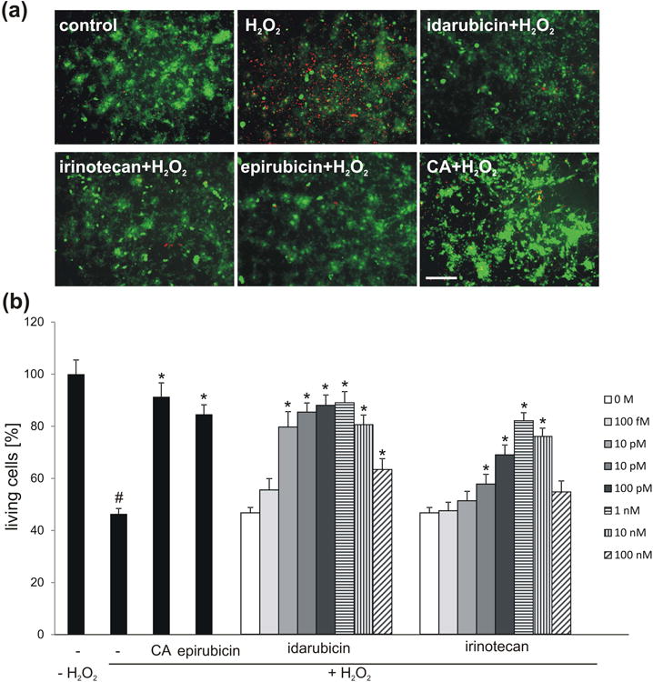
Fig. 3.

Idarubicin and irinotecan enhance survival of cerebellar granule cells in a concentration-dependent manner. (a) Representative images of cerebellar granule cells treated with 1 nM idarubicin, irinotecan or epirubicin or 30 μg/ml colominic acid (CA) and subsequently stressed using hydrogen peroxide (H2O2), and stained with calcein-AM (green) and propidium iodide (red). (b) Bar diagram shows the relative number of live neurons (mean + SEM) treated with different concentrations of irdarubicin and irinotecan, or treated with 1 nM epirubicin and 30 μg/ml CA as positive controls and stressed with H2O2. The experiment was performed independently five times with three biological replicates in each experiment (n = 5). The hash sign shows significant difference between the unstressed group (-; - H2O2) and stressed group (-; + H2O2). Asterisks signify statistically significant differences between the stressed group (-; + H2O2) and stressed and compound treated groups (epirubicin/CA/idarubicin/irinotecan; + H2O2) as determined by one-way ANOVA with Holm-Sidak post hoc test#p< 0.0001; *p< 0.005). Scale bar, 50 μm.
